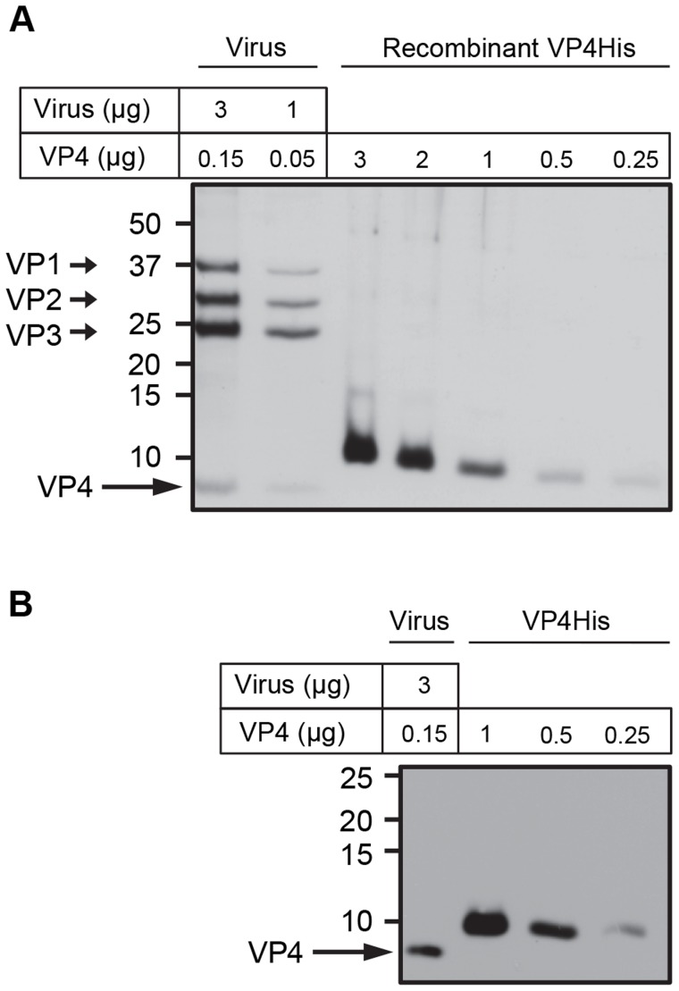
Figure 1. Purity and concentration of recombinant VP4 assessed by SDS-PAGE.
Concentration of purified VP4His estimated by protein assay was confirmed by comparison with known quantities of native VP4 in preparations of purified virus. HRV16 (3 or 1 µg, equivalent to 0.15 or 0.05 µg VP4 respectively) and VP4His at amounts indicated, were subjected to SDS-PAGE and visualized by silver staining (A) or western blot using antisera to VP4 (B). Molecular mass markers (in kilodaltons) are indicated on the left. Arrows show expected position of the indicated viral proteins. The migration of VP4His appears slower with increasing concentration as a result of the increasing concentration of DMSO in these samples. The migration of VP4His was not altered when diluted in a constant concentration of DMSO (figure S1).

