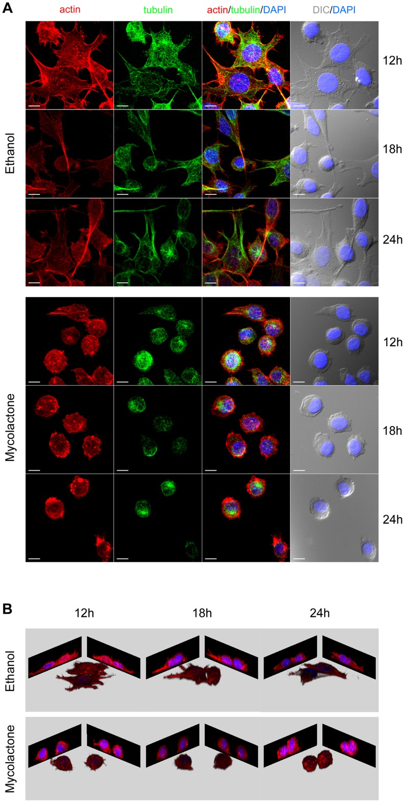
Figure 2. Mycolactone induces cytoskeletal alteration, cell round up and detachment.
Mouse fibroblasts L929 cells seeded on coverslips were incubated for 12, 18 or 24/mL of mycolactone. Cytoskeletal changes were visualized by immunofluorescence microscopy using rhodamine-phalloidin conjugate (red) and a tubulin-specific antibody (green). Nuclei were stained with 4′,6-diamidino-2-phenylindole (DAPI). The cellular shape was visualized by differential interference contrast (DIC) microscopy. White horizontal bar represents a 10 µm scale (A). Blue (nucleus) and red (actin) channels were used for 3D remodeling of the respective confocal z stacks (B).

