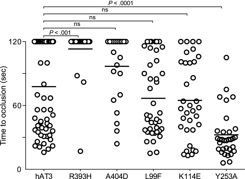
Figure 7.
Effect of human AT3 substitutions in vivo. One-cell embryos were injected with individual wild-type or mutant human AT3 cDNAs, under regulation of the ubi promoter. At 3 dpf, the endothelium was targeted and the time to occlusion measured by a blinded observer. All larvae were derived from at3+/Δ90 incrosses or at3+/Δ90 × at3Δ90/Δ90. Data from at3Δ90/Δ90 mutants injected with wild-type human AT3 cDNA were compared with at3Δ90/Δ90 injected with mutant human AT3 cDNAs, R393H (at3Δ90/Δ90, n = 25), A404D (at3Δ90/Δ90, n = 23), L99F (at3Δ90/Δ90, n = 42), K114E (at3Δ90/Δ90, n = 14), Y253A (at3Δ90/Δ90, n = 32). For statistical analysis, data were normalized to heterozygous clutchmates and significance determined by the Mann-Whitney U test followed by Bonferroni correction (see “Methods”). Horizontal bars represent the median of time to occlusion. ns, nonsignificant.

