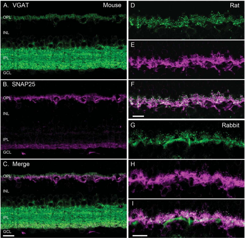
Figure 1.

SNAP25 co-localized with VGAT in horizontal cell processes. VGAT antibody staining (green) of a vertical section of mouse (A), rat (D), and rabbit (G) retinae showed immunolabeling in the outer plexiform layer (OPL) and the inner plexiform layer (IPL), and around cell bodies of the proximal inner nuclear layer (INL). SNAP25 antibody labeling (magenta) of the same section produced immunolabeling in the OPL and the proximal IPL of mouse (B), and OPL of rat (E), and rabbit (H) retinae. Merged image of the VGAT and SNAP25 immunolabeling (white) indicated co-localization of SNAP25 with VGAT in the tips of horizontal cells in mouse (C), rat (F), and rabbit (I). GCL, ganglion cell layer. Scale bar = 10 μm in C (applies to A–C), F (applies to D–F), and I (applies to G–I).
