Figure 2.

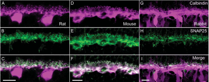
SNAP25 immunoreactivity co-localizes with that of calbindin, a horizontal cell marker. Top row shows the immunolabeling for calbindin (magenta) in (A) rat, (D) mouse, and (G) rabbit OPL. In all three species, calbindin immunoreactivity identified horizontal cell bodies, processes, and endings. In rabbit retina, calbindin immunolabeled also a subset of bipolar cells (Massey and Mills, 1996). Middle row shows the immunolabeling for SNAP25 (green) in (B) rat, (E) mouse, and (H) rabbit OPL. The SNAP25 immunoreactivity was present primarily in the tips and processes in the OPL. Bottom row shows the merged images of calbindin and SNAP25 immunreactivities (white), which demonstrated the strong expression of SNAP25 in the processes and tips of horizontal cells of all three species (C,F,I). In rabbit OPL (I) in particular, the concentration of SNAP25 in horizontal cell processes adjacent to the base of cone pedicles is evident, in addition to the tips invaginating into rod spherules. Scale bar = 10 μm in C (applies to A–C), F (applies to D–F), and I (applies to G–I).
