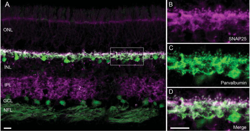
Figure 3.

SNAP25 immunoreactivity co-localized with that of parvalbumin, a horizontal cell marker in primate retina. A: A vertical section of macaque retina was double labeled with antibodies to SNAP25 (magenta) and parvalbumin (green), showing SNAP25 (SySy cl. 71.1) immunoreactivity primarily in the two plexiform layers. B–D: Higher power view of the boxed area of the outer plexiform layer (OPL) in A. B: SNAP25 immunolabeling (magenta) in processes and tips in the OPL. C: Parvalbumin immunolabeling (green) of the same region showed the presence of horizontal cell processes and tips. D: Merged image of the two immunoreactivities (white) indicated co-localization of SNAP25 to horizontal cell processes and tips in macaque retina. ONL, outer nuclear layer; INL, inner nuclear layer; IPL, inner plexiform layer; GCL, ganglion cell layer; NFL, nerve fiber layer. Scale bar = 10 μm in A and D (applies to B–D).
