Figure 5.

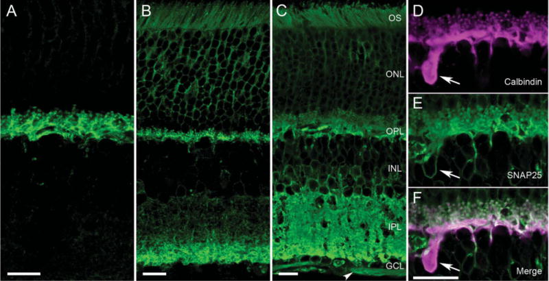
Different SNAP25 antibodies produced different immunolabeling patterns in mammalian retina. A–C: Consistently, all SNAP25 antibodies (green) appeared to immunolabel horizontal cell processes in mammalian retina (see A, mouse vertical section, Millipore/Chemicon goat Ab). Other antibodies produced labeling of the horizontal cells in the OPL and the proximal layer of the IPL (Fig. 1A). B: Still other antibodies exhibited labeling of the two plexiform layers, with the deeper layers of the IPL more strongly labeled in a vertical section of rat retina (StressGen Ab). C: SMI 81mAb produced strong labeling of both plexiform layers in mouse retina, as well as outlines of cell bodies in the ONL, INL, and GCL and the nerve fiber layer (NFL, arrowhead). D–F: These panels depict the double labeling of mouse OPL with (D, magenta) calbindin and (E, green) SMI 81 SNAP25 antibodies obtained with an aliquot of SMI 81 used in Brandstätter et al. (1996) and Morgans and Brandstätter (2000). The merged image in F indicates the co-localization of SNAP25 to horizontal cell processes, tips and soma, as well as photoreceptor terminals. OS, outer segments; ONL, outer nuclear layer; OPL, outer plexiform layer; INL, inner nuclear layer; IPL, inner plexiform layer; GCL, ganglion cell layer. Scale bar = 10 μm in A–C; 10 μm in F (applies to D–F).
