Figure 6.
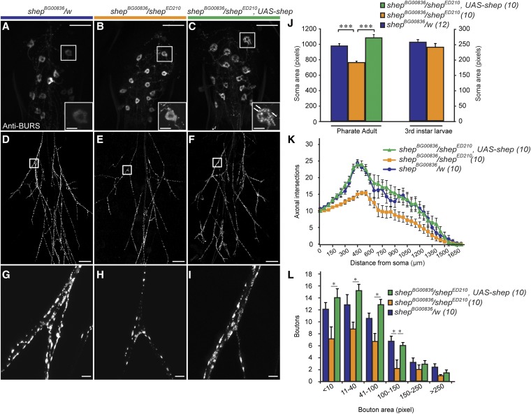
Loss of shep in shepBG00836/shepED210 animals reduced the soma area, branching of peripheral axons, and the bouton size distribution of bursicon neurons at the P14 pharate adult stage. (A–C) Immunostaining with anti-BURS antibodies showed the morphology of bursicon neurons in the abdominal ganglia. The most anterior cell on the right side is shown in the insets, with punctate peptide accumulation in the shepBG00836/shepED210, UAS–shep cell labeled by arrows. (D–F) Reduced branching of the bursicon neuron axons in the peripheral arbor in a shepBG00836/shepED210 pharate adult (E) and rescue of branching in a shepBG00836/shepED210, UAS–shep animal (F). (G–I) A reduction in bouton sizes in shep mutant animals (H) and rescue after targeted expression of shep (I) was also observed in the peripheral axon arbor. Scale bars, 50 µm (insets in A–C, 10 µm). (J) Quantification of bursicon neuron soma areas for P14 pharate adults and wandering third-instar larvae. We performed a one-way ANOVA (P < 0.000001, Tukey HSD post hoc, ***, P < 0.001) for the pharate adult values and a Student’s t-test (P = 0.934) for the wandering third-instar larval values. (K) Results of Sholl analysis of branches in the peripheral axon arbor. The space between each of the concentric rings used to count intersecting axons was 50 μm. (L) Counts of boutons along all axons 50 µm proximal and distal to the first branch of the Ab2Nv nerve (squares). (*) P < 0.05 (two-way ANOVA, P < 0.00001; Tukey HSD post hoc test. Scale bars, 100 µm.

