
Figure 5.
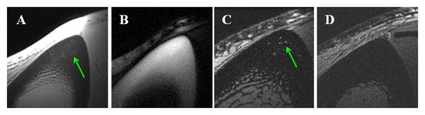
Axial imaging of the tibia mid-shaft of a 58 year old healthy volunteer with UTE (A), IR-UTE (B) and FSE (C) sequences, and FSE imaging of a 39 year old healthy volunteer (D). UTE detects signal from bone but with limited contrast due to much higher signal from the surrounding muscle and bone marrow fat (A). IR-UTE shows high signal and contrast for cortical bone with excellent suppression of signals from the surrounding muscle and bone marrow fat (B). The fine structures in FSE images correspond to the large Haversian canals (C). The younger volunteer shows no structure in cortical bone with the FSE sequence, consistent with bone without larger canals (D).