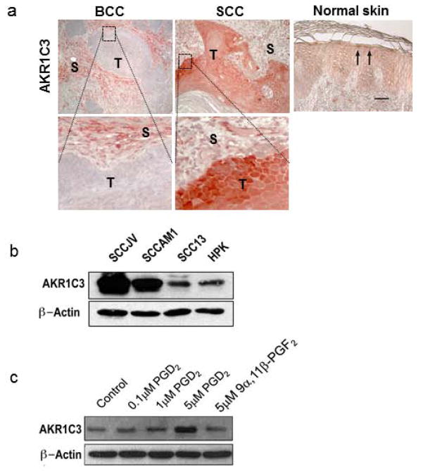
Figure 1. AKR1C3 overexpression in tumor mass of SCC and its induction by PGD2.
(a) AKR1C3 expression was evaluated in six cases of SCC, seven cases of BCC and six cases of normal buttock skin by immunohistochemical staining. Representative figures demonstrating AKR1C3 expression in normal skin and within the tumor mass (T) and tumor stroma (S) of SCC and BCC. Strong immunoreactivity was observed in SCC while undetected in BCC. Normal skin showed moderate expression mainly in suprabasal layers (arrows). Bar=30μM. (b) expression of AKR1C3 in cell lines SCCAM1, SCCJV and SCC13. To evaluate the expression of AKR1C3 in the different SCC cell lines, cells cultured at confluence for 24 hours following by protein extraction and western blot analysis (n=3; representative blot is shown). (c) PGD2-induced AKR1C3 expression in SCC cells. SCC cells were treated for 24 hours with various doses of PGD2 or its AKR1C3-mediated metabolite 9α11βPGF2 as indicated and AKR1C3 expression was assessed by western blot. A representative experiment in which SCCAM1 was used is shown.

