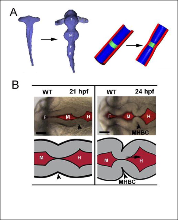
Fig 2. Formation of primary vesicles in brain tube.
(A) Chick embryo. Reconstructed brain lumen at stages 10 and 12 (left) and schematic of boundary formation (right). Boundaries between vesicles are created by circumferential actomyosin contraction (green region) at apical side of wall. From [47*]. (B) Zebrafish embryo. Boundary between midbrain and hindbrain (arrowhead) forms in two steps: radial shortening (left) and basal constriction (right) of neuroepithelial cells. (F= forebrain; M = midbrain; H = hindbrain; MHBC = midbrain-hindbrain boundary constriction) From [48*].

