
Fig. 2. Chromatin diminution in Parascaris and Ascaris.
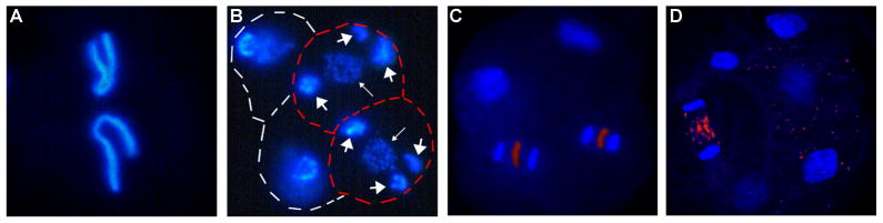
A and B. P. univalens embryos. A. 1-cell embryo showing the single pair of germline chromosomes. B. 4-cell embryo with two cells (outlined in red) undergoing diminution. The retained portions of the germline chromosomes are fragmented into many smaller chromosomes (small arrows). The heterochromatic arms that will be eliminated (big arrows) remain visible. C and D. A. suum embryos. C. 4-cell embryo with two cells undergoing chromatin diminution. D. 6-cell embryo with one cell undergoing chromatin diminution. Note that DNA to be eliminated is present as fragments (artificially colored red) between chromosomes segregating in early anaphase (C); DNA fragments (red) derived from a previous cell diminution can be seen in the cytoplasm of cells to the right (D).