
Figure 1.
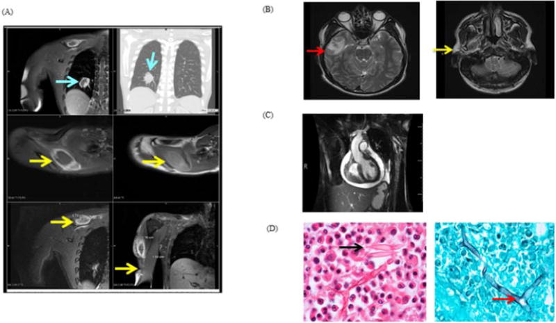
A, Chest MRI. Lesions are visible in the right lung (top panels), right supraspinatus (arrows, middle panels) and deltoid (lower panels); B, Brain MRI T2W images. Thick-walled right temporal lobe lesion (left); enhancement of the right muscles of mastication (right); C, Cardiac MRI: Pericardial effusion with mild RV collapse; D, H&E inguinal lymph node biopsy (left) with intense eosinophilic infiltrate, Charcot-Leyden crystals (arrow); silver stain of deltoid muscle (right). Aseptate fungal elements with 90° branching hyphae were observed (arrow).