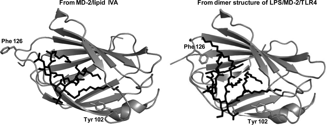
Fig. 6. Ribbon models of MD-2 bound with lipid IVA (36) or LPS (37) based on X-ray crystal structure.
The structure of MD-2 bound to LPS is based on LPS.MD-2.TLR4ECD with TLR4 not displayed “. The MD-2 is shown in gray while the ligand is shown in black. Note the different orientation of Phe126 in the two complexes. Note also the position of the side chain of Tyr102 within the hydrophobic pocket of MD-2.

