Figure 3.
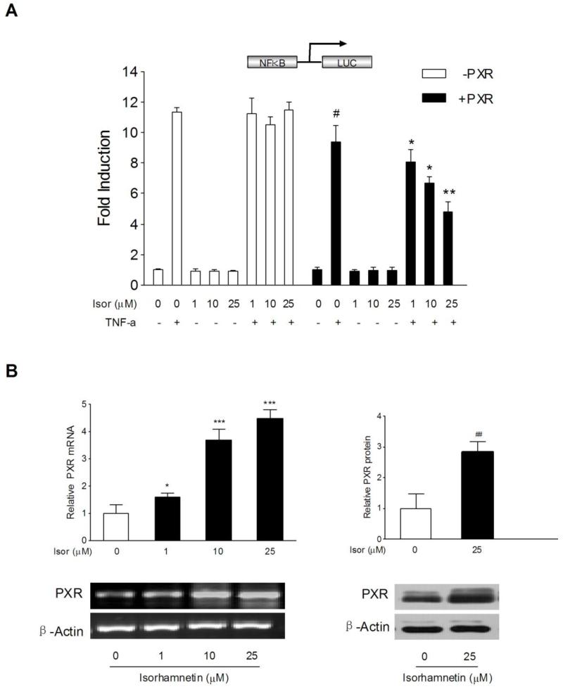
Role of PXR on isorhamnetin-mediated NF-κB luciferase inhibition and the effects of isorhamnetin on PXR expression. (A) HT-29 cells were electroporated with the pGL4.32[luc2P/NF-κB-RE/Hygro] reporter alone or coelectroporated with pSG5-hPXR and pRL-TK. After transfection, cells were treated either with TNF-α (20 ng/ml) alone or cotreated with isorhamnetin (0, 1, 10, 25 μM) for 24 h. Luciferase activity was determined with cell lysates, and the results are expressed as fold induction of control cells (designated as 1). Data are presented as the mean ± SD of three independent experiments. # P < 0.05 vs. TNF-α alone-treated samples without hPXR transfection; *p < 0.05, **P < 0.01 vs. TNF-α alone-treated samples with hPXR transfection. (B) The mRNA expression of PXR (n=3) was assessed by semi-quantitative RT-PCR in LS174T cells treated with isorhamnetin (0, 1, 10, 25 μM); the protein expression of PXR (n=3) was determined by western blot in LS174T cells treated with isorhamnetin (25 μM). *p < 0.05, ** p < 0.01, ***P < 0.001 vs. vehicle-treated group.

