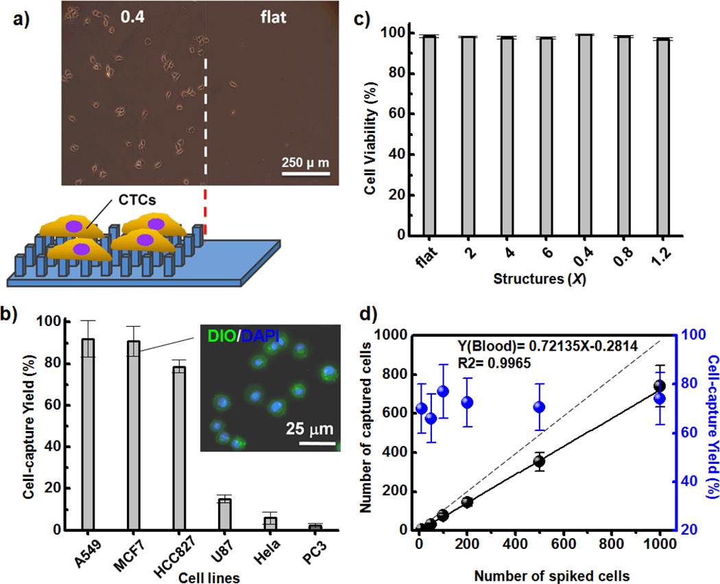
Figure 4.
a) Microscopy image of MCF7 cells captured on (left) patterned PEDOTAc-0.4 and (right) the flat substrate. b) Cell-capture efficiencies from suspensions of breast (MCF), lung (A549, HCC827), cervical (HeLa), prostate (PC3), and brain (U87) cell lines; inset: two-color fluorescence image based on DiO membrane (green) and DAPI nuclear staining, used to identify the captured morphology of MCF7 cells on PEDOTAc-0.4. c) Quantitative evaluation of cell viability of MCF7 cells captured on various PEDOTAc structures; error bar represents the standard deviation from three repeats. d) Capture efficiencies of various contents of MCF7 cells in whole blood; dashed line: ideal cell-capture yield when the various spiked cell densities were greater than 100%.

