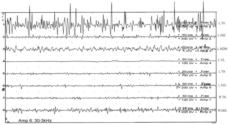
Figure 2.
Subject 30 is now standing and leaning forward on a chair, taking much of his weight off his legs. He asserts his legs feel, “steady and secure” and the “little, unsteady movements and jerks” within them have utterly, subjectively abated. The SEMG tracing lasting one second reveals high amplitude, tonic motor activity in his left triceps and only very low amplitude, tonic activity in his legs.
L= left; R= right; Tri= triceps; WE= wrist extensors; ADM= abductor digiti minimi; VL= vastus lateralis; TA= tibialis anterior; MG= medial gastrocnemius; ms= milliseconds, uV= microvolts

