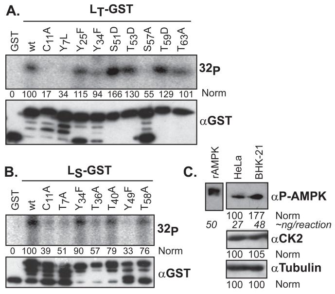
Figure 2.
LX phosphorylation in cytosol. LT-GST (A) or LS-GST (B) proteins were incubated with HeLa cytosol and [γ-32P]-ATP. After GST extraction and SDS-PAGE, the proteins were detected by autoradiography (32P), or Western analysis (αGST). After densitometry, the signals were normalized to the GST control (“0”) and wt (“100”) samples. The uppermost αGST band is the fusion protein. (C) Equivalent cytosol samples (HeLa, BHK) to A and B, were probed by Western analysis for AMPK, CK2 and tubulin signals, relative to a standardized sample of rAMPK.

