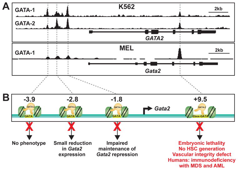
Figure 1.
Distinct functional properties of Gata2 GATA switch sites. (A) ChIP-seq profiles of GATA-2 chromatin occupancy at GATA2 locus in model erythroid cell systems: human K562 and mouse MEL cells. GATA-2 occupies established GATA factor binding regions termed GATA switch sites, which contain evolutionarily conserved GATA motifs and multiple attributes of transcriptional enhancers. (B) Schematic showing the GATA switch sites (−3.9, −2.8, −1.8 and +9.5 kb upstream of the 1S promoter transcription start site), and phenotypes resulting from deletion of the respective sites. The red X mark denotes the cis-element deletion from the mouse genome.

