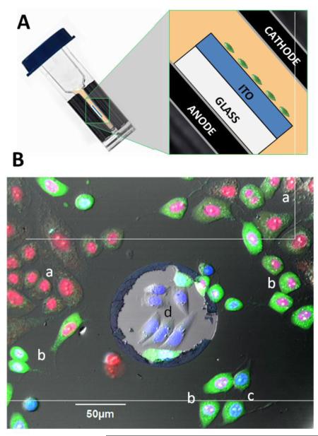
Fig. 1.
Exposure of ITO-coated coverslips in electroporation cuvettes (A) and the critical role of the ITO layer in delivering the electric field to adherent cells (B). The schematic in (A) shows the position of the ITO coverslip with cells inside an electroporation cuvette (not to scale). (B): Overlaid fluorescence and DIC images of CHO cells at 2 hr after nsPEF exposure (20 pulses, 300 ns duration, 20 Hz, 600 V, on ITO coverslip in a 1-mm electroporation cuvette). The cells were held in 2 mM Ca2+ buffer with Hoechst (blue), Yo-PRO-1 (green), and Pr (red), see Methods for details. The lighter area in the center is a random defect in ITO coating. Labels indicate: (a) dead cells with Pr-stained nuclei; (b) dying cells with Pr-stained nucleus and Yo-PRO-1 stained cytosol; (c) electropermeabilized but otherwise healthy cells with YO-PRO-1 staining of cytosol and mixed Hoechst and Yo-PRO-1 staining of the nucleus; and (d) healthy, unaffected cells displaying only Hoechst labeling of the nucleus. Note that the unaffected cells are only found within the ITO defect.

