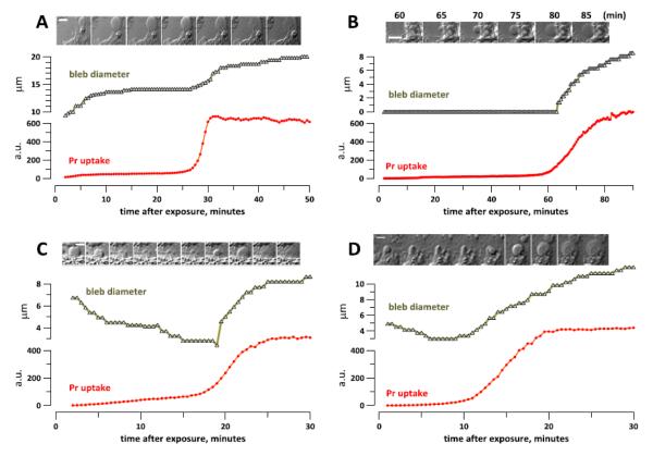
Fig. 6.
Bleb growth in nanoporated cells accelerates or re-starts with the abrupt Pr uptake in BPAE (A-C) and CHO cells (D). After nsPEF exposure (20 pulses, 300 ns duration, 20 Hz, 600 V, on ITO coverslip in a 1-mm electroporation cuvette), cells were incubated in the 2 mM Ca2+ buffer (A) or in 2 mM Ca2++sucrose buffer (B-D). The insets show DIC images of the measured blebs at the timepoints which correspond the X axis (A,C,D). In B, the time when images were taken is indicated in the legend. The calibration bar is 10 μm (A) or 5 μm (B-D).

