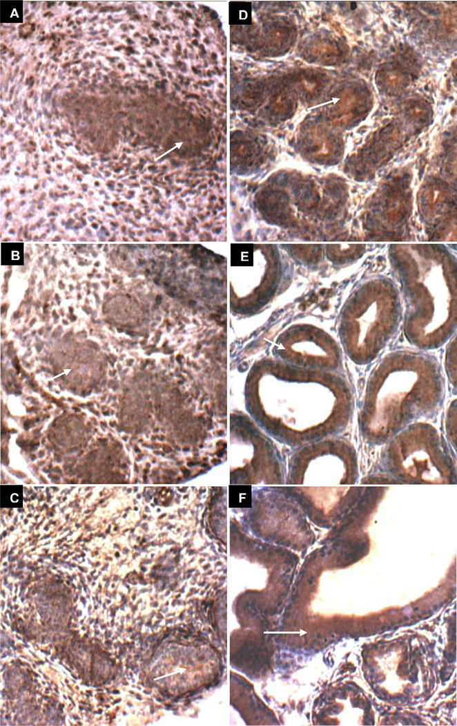
Figure 4. GALNS immunohistochemical staining of prostate tissue in oil control rats on Days 1–30.
A-F. GALNS immunohistochemistry on days 1(A), 3(B), 6(C), 10(D), 15(E), and 30(F) demonstrates increasing definition of brown immunostaining of GALNS in the developing epithelial areas, surrounding an evolving lumen and distinct from the relatively non-staining (blue) stroma. White arrows denote representative sites of positive brown cytoplasmic staining of epithelial cells in developing acini. [GALNS=galactose-6-sulfatase]

