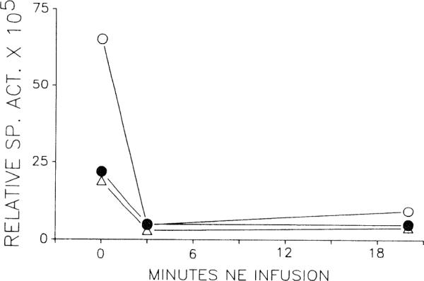
FIG. 3.

Relative specific activity of venous adenosine after selective endothelial labeling with [3H]adenosine (n = 3). Time zero represents resting conditions. Specific activity falls and remains low during NE infusion. Each symbol represents one heart.
