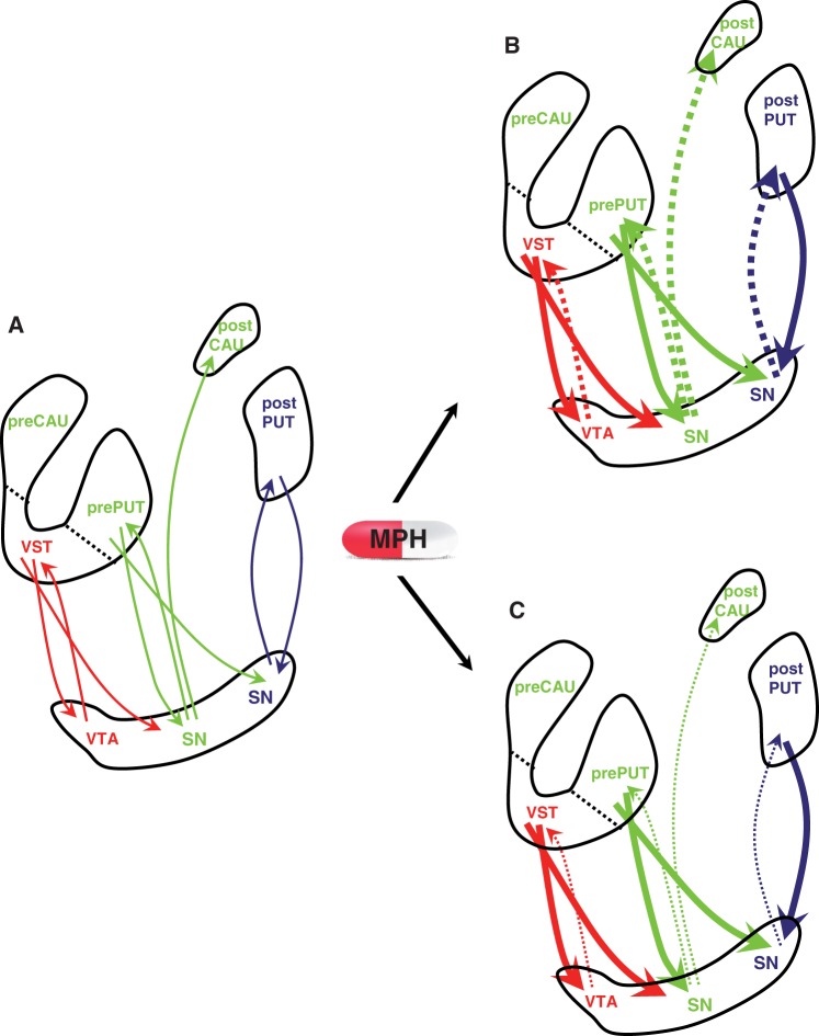
Figure 12.
Schematic model of dopamine activity along striato-nigro-striatal circuits at baseline (A) and after oral methylphenidate (MPH) in high (B) and low performers (C). Ascending arrows represent projections from dopamine (DA) neurons located in the substantia nigra (SN)/ventral tegmental area (VTA) to the striatum and descending arrows represent GABA-ergic projections from the striatum to the substantia nigra/ventral tegmental area. Different colours represent different striatal functional circuits (red = limbic, green = associative, blue = sensorimotor). Methylphenidate administration (B) increased postsynaptic catecholamine stimulation (solid lines), leading to inhibition of dopamine neuron firing (dashed lines) by activation of synthesis- and release-regulating dopamine autoreceptors. It is suggested that the level of responsivity of dopamine neurons and subsequent striato-nigro-striatal neuromodulation following methylphenidate administration was dependent on baseline dopamine activity in the striatum. Adapted from Haber et al. (2000) and Martinez et al. (2003). CAU = caudate PUT = putamen; VST = ventral striatum.

