
Figure 1.
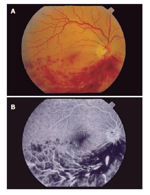
A: Fundus photograph of the right eye with extensive intraretinal hemorrhages and cotton-wool spots in the inferonasal and inferotemporal regions; B: Fluorescein angiography with segmental hypoperfusion, dilation and tortuosity of the retinal veins (arrow), compatible with inferior branch retinal vein thrombosis.