
Figure 3.
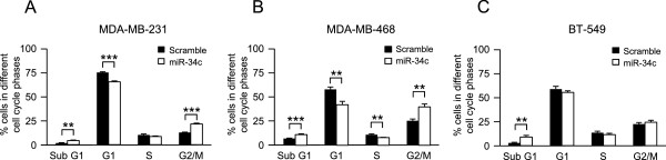
Effect of miR-34c on cell cycle distribution of breast cancer cells. Following transfection of MDA-MB-231 (A), MDA-MB-468 (B) and BT-549 (C) breast cancer cells with miR-34c mimic or negative control for 96 h, nuclei were stained with propidium iodide solution and analyzed for DNA content by flow cytometry. Data (mean ± SEM, n = 5) represent percentage cells in different phases of the cell cycle with miR-34c related to scramble treatment. Asterisks indicate statistically significant differences (* p < 0.05, ** p < 0.01, *** p < 0.001, Student’s t-test) compared to control cells.