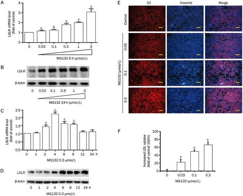
Figure 1.
Effects of MG132 on LDLR expression in and LDL uptake by HepG2 cells. (A) Real-time quantification of the LDLR mRNA level and (B) Western blot analysis of LDLR protein in HepG2 cells treated with MG132 (0.03, 0.1, 0.3, 1, and 3 μmol/L) for 6 h. Time-course of the LDLR mRNA (C) and protein (D) levels in HepG2 cells exposed to MG132 (0.3 μmol/L) for 24 h. DiI-LDL uptake was assessed in cells treated with the indicated concentrations of MG132 for 24 h; (E) Representative images of cells associated with DiI-LDL (Red) and Hoechst-stained nuclei (Blue). Scale bar 50 μm; (F) The normalized fluorescence of isopropanol-extracted DiI (520 and 570 nm). Data are presented as the mean±SEM of at least three independent experiments. bP<0.05, cP<0.01 vs the vehicle-treated groups.

