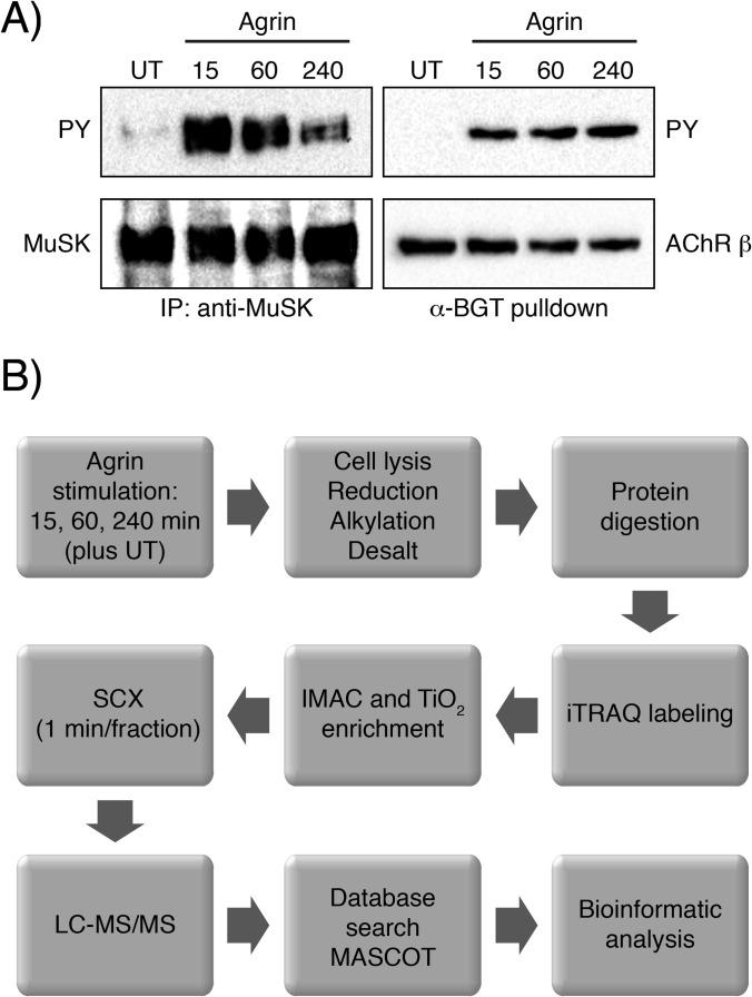
Fig. 1.
MuSK and AChR β phosphorylation in muscle cells and MS experimental strategy. A, cell lysates from agrin-induced muscle cells were subjected to an immunoprecipitation with anti-MuSK antibodies or a pulldown using biotinylated α-BGT. Samples were analyzed by immunoblotting using antibodies against phosphotyrosine, MuSK or AChR. B, experimental strategy for phosphoproteomic analysis. Muscle cells unstimulated (UT) or stimulated across three time points were lysed. Proteins were extracted, digested and isotopically labeled with the iTRAQ 4-plex reagent. Global phosphorylation was assessed via TiO2 and IMAC enrichment. Eluted peptides were then analyzed via LC-MS/MS and the resulting data were processed for phosphopeptide identification and quantification as described in “Experimental Procedures.”

