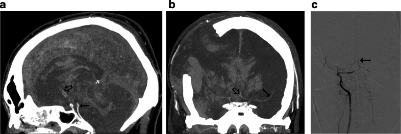
Fig. 3.
CTA findings in a 50-year-old man (patient no. 45) with traumatic brain injury (epidural hematoma in the right parietal region, massive intracerebral, and subarachnoid and intraventricular hemorrhage) and right sided craniectomy presented with signs of BD on clinical examination: a Ten millimeter maximum intensity projection (MIP) in sagittal plane. CTA shows opacification of the BA (thin arrow) and a trace of contrast in A2 segments of the ACAs (thick arrow). b Ten millimeter MIP in coronal plane. CTA shows opacification of the M1 segment of the left MCA (thin arrow) and the A1 segments of the ACAs (thick arrow); these findings exclude the diagnosis of BD according to the 10-point scale but confirm BD according to the 7- and 4-point scales. c Catheter angiography of the right VA performed 0.5 h later revealed delayed, residual filling of the BA (arrow) that occurred 21 s after injection. This result was interpreted as stasis filling consistent with the diagnosis of BD

