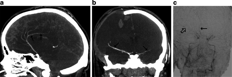
Fig. 4.
CTA findings in a 22-year-old woman (patient no. 42) with a brain stem ischemic stroke and a right sided craniectomy who presented with signs of BD on clinical examination. a Ten millimeter MIP in sagittal plane. CTA shows opacification of the right pericallosal artery (thin arrows); b Ten millimeter MIP in coronal plane. CTA shows opacification of the M1 segments of the MCAs (thin arrows). These findings exclude the diagnosis of BD according to the 10- and 7-point scales but confirm BD according to the 4-point scale. c Catheter angiography from the aortic arch performed 1 h later revealed delayed, residual filling of the M1 segment of the right MCA (thick arrow) and A2 segment of the right ACA (thin arrow) that occurred 32 s after injection. This result was interpreted as stasis filling consistent with the diagnosis of BD

