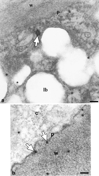
Fig. 6.

EM immunogold identification of cutinsomes in O. umbellatum ovary epidermal cell. a Labelled cutinsome (white arrows) probably leaving the lipotubuloid. b Cutinsomes (white arrows) out of the cytoplasm at the other side of the plasmalemma and in the cell wall. c cytoplasm, lb lipid bodies, p plasmalemma, w cell wall. Scale bar, 100 nm
