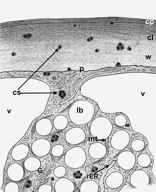
Fig. 9.

Scheme of cutinsome localization in O. umbellatum ovary epidermal cell. The cutinsomes form in a lipotubuloid and then probably move towards the cell wall and cuticle through the plasmalemma (on the basis of enclosed electron microscopy–immunogold technique images). cl cuticular layer, cp cuticle proper, cs cutinsomes, G Golgi apparatus, lb lipid bodies, mt microtubules, p plasmalemma, rER rough endoplasmic reticulum, v vacuole, w cell wall
