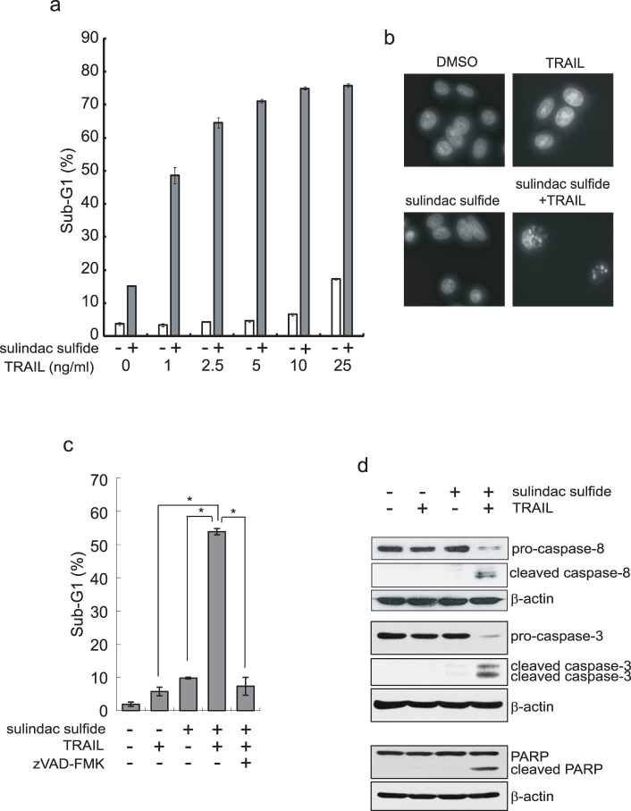
Figure 1. Sulindac sulfide enhanced TRAIL-induced apoptosis in SW480 cells.
(a) SW480 cells were treated with 200 μM sulindac sulfide and/or the indicated concentrations of TRAIL for 24 h. Cells were analyzed for DNA content by PI staining (FL2-H) using a flow cytometer. The percentages of sub-G1 are shown as a bar graph. (b) DAPI staining of SW480 cells. SW480 cells were treated with 200 μM sulindac sulfide and/or 10 ng/ml TRAIL for 24 h. Nuclear morphology was visualized using DAPI staining under a fluorescence microscope. (c) SW480 cells were treated with 200 μM sulindac sulfide and/or 10 ng/ml TRAIL with or without 20 μM zVAD-fmk for 24 h. The effects were analyzed as described in (a). Data represent the means +/− S.D. of three determinations. *: p < 0.05 (d) Western blotting for caspase-8, caspase-3 or PARP. SW480 cells were treated with 200 μM sulindac sulfide and/or 10 ng/ml TRAIL for 24 h. β-actin was used as a loading control.

