Figure 5.
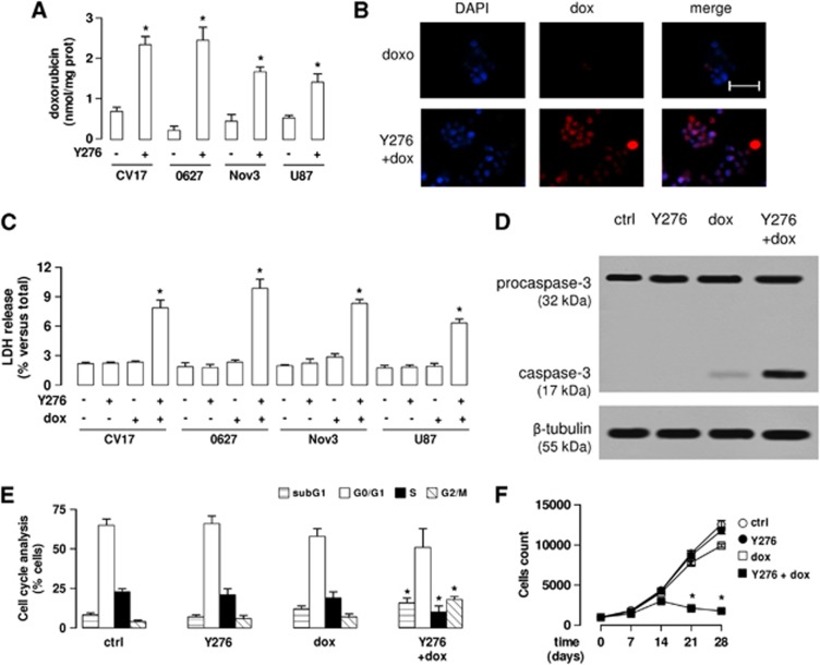
The RhoA kinase (RhoAK) inhibitor Y27632 increases the doxorubicin delivery and cytotoxicity in glioblastoma cells co-cultured with blood–brain barrier cells. The hCMEC/D3 cells were grown for 7 days up to confluence in Transwell inserts; CV17, 01010627, Nov3, and U87-MG cells were seeded at day 4 in the lower chamber. After 3 days of co-culture, the supernatant in the upper chamber was replaced with fresh medium without (− or ctrl) or with Y27632 (Y276; 10 μmol/L for 3 hours). After this incubation time, doxorubicin (dox; 5 μmol/L) was added in the upper chamber for 3 hours (panels A and B) or 24 hours (panels C–F), then the following investigations were performed. (A) Fluorimetric quantification of intracellular doxorubicin in glioblastoma cells. Data are presented as means±s.d. (n=4). Versus untreated (−) cells: *P<0.001. (B) The 01010627 cells were seeded on sterile glass coverslips, treated as reported above, then stained with 4′,6-diamidino-2-phenylindole dihydrochloride (DAPI) and analyzed by fluorescence microscopy to detect the intracellular accumulation of doxorubicin. Magnification: × 63 objective (1.4 numerical aperture); × 10 ocular lens. The micrographs are representative of three experiments with similar results. Scale bar, 20 μm. (C) The glioblastoma cells were checked spectrophotometrically for the extracellular release of lactate dehydrogenase (LDH) activity. Data are presented as means±s.d. (n=4). Versus untreated (−) cells: *P<0.001. (D) The whole-cell lysates from 01010627 cells were resolved by SDS–PAGE and immunoblotted with an anti-caspase 3 antibody (recognizing both pro-caspase and cleaved active caspase). The β-tubulin expression was used as a control of equal protein loading. The figure is representative of three experiments with similar results. (E) Cell cycle analysis. The distribution of the 01010627 cells in sub-G1, G0/G1, S, G2/M phase was analyzed by flow cytometry, as detailed under Materials and Methods. Data are presented as means±s.d. (n=4). Versus ctrl: *P<0.005. (F). After 3 days of co-culture between hCMEC/D3 and 01010627 cells, the medium of the upper chamber was replaced with fresh medium (open circles) or medium containing Y27632 (Y276; 10 μmol/L for 3 hours, solid circles), doxorubicin (dox; 5 μmol/L for 24 hours, open squares), Y27632 (Y276; 10 μmol/L for 3 hours) followed by doxorubicin (doxo; 5 μmol/L for 24 hours, solid squares). Drug treatments were repeated every 7 days, as reported in the Materials and Methods section. The proliferation of glioblastoma cells was monitored weekly by crystal violet staining. Measurements were performed in triplicate and data are presented as means±s.d. (n=4). Versus ctrl: *P<0.001.

