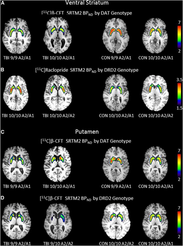
Figure 2.
Images depict regional binding for [11C]β-CFT and [11C]raclopride based on SRTM2 analysis using cerebellum as reference region for a single slice through the ventral striatum (A/B) and putamen (C/D) in the axial plane overlaid onto high resolution anatomic MRI. (A) Shows a representative image of the ventral striatum and DAT binding in persons with DAT1 genotype 9/9 and DAT1 genotype 10/10 while keeping the DRD2 constant for the A2/A1 genotype in TBI (left two panels) and controls (right two panels). Panel B shows a representative image through the ventral striatum of DRD2 binding in persons with DRD2 genotype A2/A1 and DRD2 genotype A2/A2 while keeping DAT constant for the 10/10 genotype in TBI (left two panels) and controls (right two panels). Panel C shows a representative image of the putamen and DAT binding in persons with DAT1 genotype 9/9 and DAT1 genotype 10/10 while keeping the DRD2 constant for the A2/A1 genotype in TBI (left two panels) and controls (right two panels). Panel D shows a representative image of the putamen and DAT binding in persons with DRD2 genotype A1/A2 and A2/A2 while keeping the DAT1 genotype constant for the 10/10 genotype in TBI (left two panels) and controls (right two panels). Note: TBI 9/10 A2/A2 scale was 1.5 to 6. CON, control; DAT, dopamine transporter; MRI, magnetic resonance imaging; TBI, traumatic brain injury.

