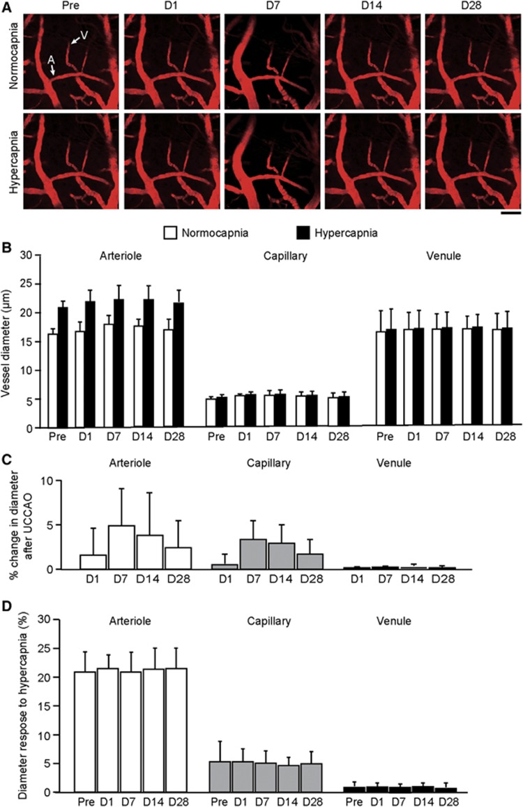
Figure 4.
(A) Longitudinal imaging of cortical vessels at the cortical surface in the hemisphere contralateral to unilateral common carotid artery occlusion (UCCAO). Scale bar, 50 μm. (B) Longitudinal diameters in cortical microvessels during normocapnia and hypercapnia in the hemisphere contralateral to UCCAO. (C) Longitudinal percentage changes in cortical microvessel diameter from the preoperative value in the hemisphere contralateral to UCCAO. Although arterioles were slightly dilated 7 days after UCCAO, statistically significant dilation was not found in any of the three microvasculature components throughout the experimental period. (D) Longitudinal vascular responses to hypercapnia in cortical microvessels in the hemisphere contralateral to UCCAO. There were no significant differences in responses throughout the experimental period. The error bars indicate standard deviation. A, arteriole; V, venule. Normocapnia (white); hypercapnia (black).

