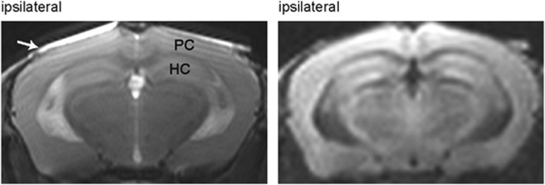
Figure 6.
Magnetic resonance imaging experiments obtained 30 days after unilateral common carotid artery occlusion. The panel on the left shows a T2-weighted image. The panel on the right shows a diffusion-weighted image. The images were acquired at the area including the parietal cortex (PC) and hippocampus (HC). No abnormal changes in signal intensity were observed. The white arrow indicates the cover glass.

