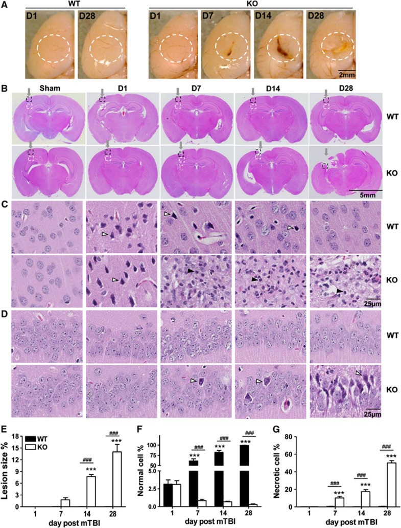
Figure 2.
Secondary brain injury occurs in KO mice but not in WT mice after gentle insult. (A) Gross morphology of the impact, left hemisphere of the brain. Note, a normal appearance on day 1 (D1) and day 28 (D28) of WT brain after trauma, but a lesion deteriorating over time in KO brain, marked by white line dash circles. (B–D) Histologic examination of coronal sections stained with hematoxylin and eosin from WT and KO brains at indicated days after mTBI. The impact site in sections panel B is pointed by an arrow; the injured neocortex is highlighted in a dash black line square and enlarged in panel C; and the hippocampus underneath is outlined by a dashed white line square and magnified in panel D. Unfilled arrows indicate one of the necrotic cells in each field and filled arrows indicate one of the infiltrated leukocytes. All data in the figures were representative of six mice per group. Percentages of a lesion size over the whole brain section in panel B were determined by Image J and expressed as means±s.e.m. in panel E. Percentages of normal cells in panel C or necrotic cells in panel D relative to a total number of cells counted in the same fields were determined as detailed in Materials and Methods and shown as means±s.e.m. in panel F and panel G, respectively. ***P<0.001 compared with Day 1 and ###P<0.001 in the presence or absence of IEX-1. IEX-1, immediate early responsive gene X-1; KO, knockout; mTBI, mild traumatic brain injury; WT, wild-type.

