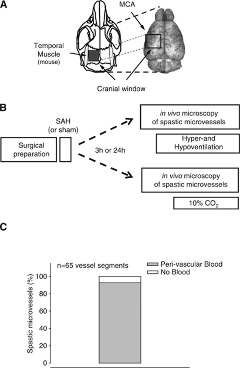
Figure 1.
(A) Schematic drawing of the rodent skull (left) and a dorsal image of the brain (right) indicating the location of the cranial window used for intravital microscopy in mice. MCA, middle cerebral artery. (B) Schematic drawing of the experimental protocol used in this study. SAH, subarachnoid hemorrhage. (C) Percentage of spastic microvessels ensheathed by blood.

