Figure 4.

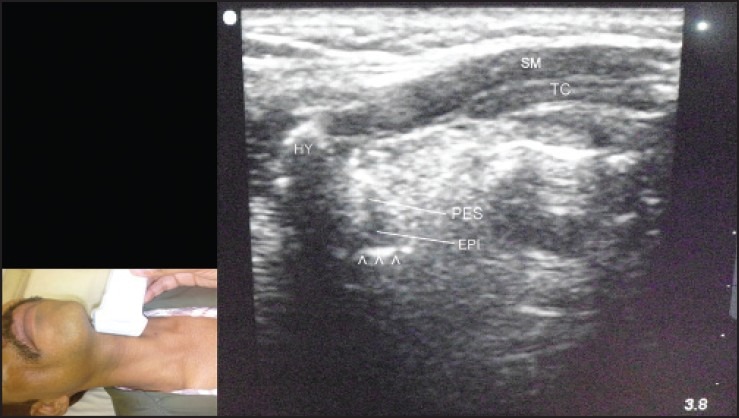
Left parasagittal view through thyrohyoid membrane using a linear transducer. The scan shows epiglottis (EPI), preepiglottic space (PES), hyoid bone (HY), strap muscles (SM), air-mucosal interface (arrowheads), and thyroid cartilage (TC)

Left parasagittal view through thyrohyoid membrane using a linear transducer. The scan shows epiglottis (EPI), preepiglottic space (PES), hyoid bone (HY), strap muscles (SM), air-mucosal interface (arrowheads), and thyroid cartilage (TC)