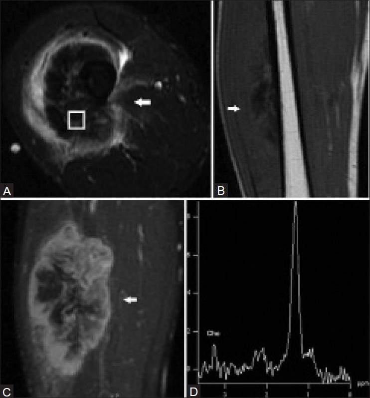
Figure 2 (A-D).

A 31 year old male with high-grade osteosarcoma of the right thigh. Axial short TI inversion recovery (STIR) fast spin-echo MRI image (A), coronal T1-weighted spin-echo MRI image (B), and coronal fat-saturated dynamic contrast-enhanced fast spin-echo MRI image acquired 40 s after contrast injection (C) demonstrate a heterogeneous enhancing mass in the anterolateral right thigh with central areas of necrosis and surrounding edema. A single-voxel MR spectroscopic map (D) demonstrates a discrete choline peak at 3.2 ppm. (Reprinted with permission from AJR)[5]
