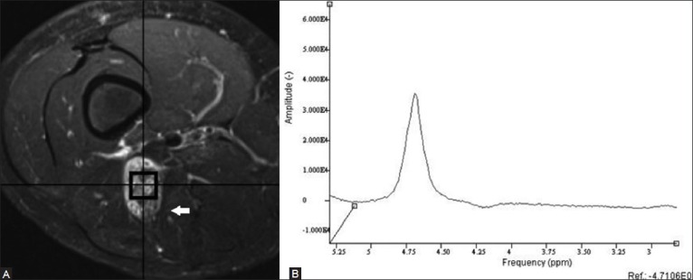
Figure 4 (A, B).

An 11 year old male with history of neurofibromatosis type 1 presents with marked enlargement of the right sciatic nerve. Axial fat-suppressed T2W fast spin-echo MRI image (A) demonstrates a mass that was found to be a benign neurofibroma. Single-voxel proton MR spectroscopic map (B) no discernible choline peak at 3.2 ppm, compatible with a benign lesion. (Reprinted with permission from AJR)[5]
