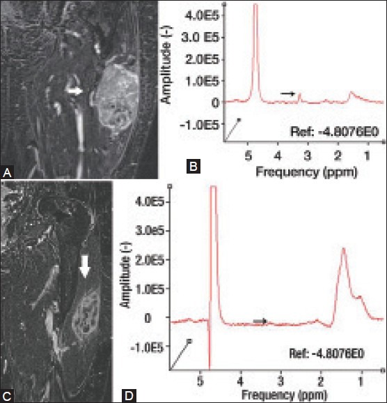
Figure 6 (A-D).

An 81 year old female with pleomorphic rhabdomyosarcoma. Coronal delayed contrast-enhanced MRI (A) and proton MRS (B) demonstrate an avidly enhancing mass with a discrete choline peak at 3.2 ppm. The patient was subsequently treated with chemotherapy. Post-treatment coronal delayed contrast-enhanced MRI (C) demonstrates substantially decreased enhancement within the lesion. Post-treatment proton MRS (D) demonstrates marked interval decrease of choline peak at 3.2 ppm, indicating chemotherapy-related tumor necrosis. (Reprinted with permission from Radiology)[2]
