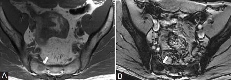
Figure 1 (A and B).

Island of red marrow in the sacrum. A 49 year old man with recurrent bloating underwent a MR enterography, which demonstrated an incidental lesion in the sacrum. He was recalled for in-phase and out-of-phase T1W MRI imaging. (A) In-phase T1W MRI image demonstrates the lesion (arrow) is slightly hyperintense to skeletal muscle (B) On the out-of-phase T1W MRI image, there is loss of signal due to the presence of intermixed fatty marrow (arrow)
