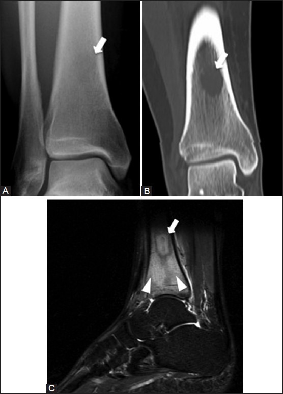
Figure 22 (A-C).

Osteomyelitis with Brodie's abscess. A 29 year old female with left lower leg pain. (A) AP radiograph of ankle demonstrates a faint radiolucency (arrow) in the distal tibial diaphysis (B) Coronal CT image shows that the lesion (arrow) is well demarcated with a non-sclerotic rim (C) Sagittal T2W fat-saturated MRI image shows the hyperintense intraosseous abscess (arrow) with surrounding marrow edema (arrowheads)
