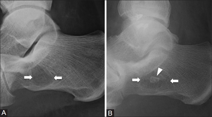
Figure 4 (A and B).

Calcaneal pseudocyst and intraosseous lipoma. (A) Lateral ankle radiograph of a 39 year old female with foot pain demonstrates a radiolucency (arrows) in the anterior calcaneus due to decrease in bony trabeculae (B) Lateral ankle radiograph of a 45 year old man with an intraosseous lipoma (arrows) shows a similar radiographic appearance to the calcaneal pseudocyst; however, there is focal central calcification (arrowhead) due to fat necrosis
