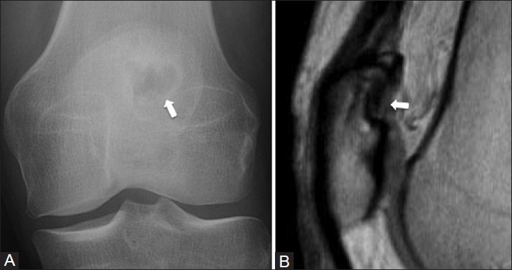
Figure 5 (A and B).

Dorsal defect of the patella. A 38 year old female with left knee pain. (A) AP radiograph of the knee demonstrates a focal radiolucency (arrow) in the superolateral aspect of patella (B) Sagittal PDW MRI image shows a focal area of cortical irregularity with intact overlying hyaline cartilage (arrow)
