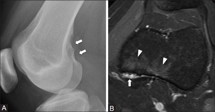
Figure 7 (A and B).

Avulsive cortical irregularity of the posterior femur. An 18 year old female with left knee pain. (A) Lateral radiograph of knee demonstrates an area of cortical irregularity at the medial aspect of the distal femoral metaphysis (arrow) (B) Corresponding axial T2W fat-saturated MRI image shows marrow edema (arrowhead) at the area of cortical irregularity (arrow)
