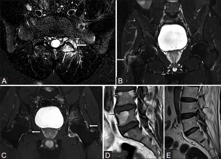
Figure 12 (A-E).

Associated findings in seronegative spondyloarthropathy-related sacroiliitis on MRI. (A) Axial STIR MR image at L5-S1 shows left facetal joint subchondral edema (white arrows). (B) Coronal STIR MR image through both hip joints shows edema at tendinous insertion in the right greater tuberosity of femur, suggestive of enthesopathy (white arrow). (C) Coronal STIR MR image through both hip joints shows minimal joint effusion with prominent synovial thickening indicating synovitis (white arrows). (D) Sagittal T1W MR image of lumbosacral spine shows bridging syndesmophyte (thin black arrow), fatty deposition at the superior aspect of anterior and posterior margins of L4 vertebral body indicating shiny corner sign (thick black arrow), and fatty deposition around L4-5 facet joint (white arrow). (E) Sagittal T1W MR image of lumbosacral spine shows ankylosis of the facet joint (black arrow). Detection of any of these findings in the imaging field in patients with sacroiliitis can support a diagnosis of seronegative spondyloarthropathy
