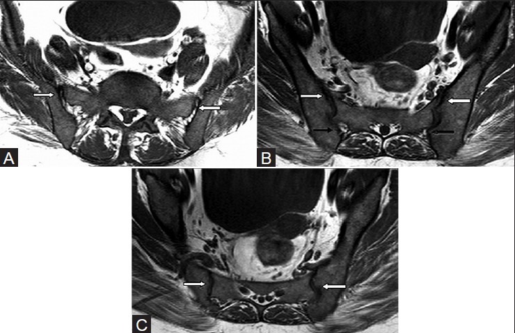
Figure 2 (A-C).

Normal anatomy of the sacroiliac joint. Axial T1W MRI images at three different levels in a 29 year male: (A) upper portion is predominantly ligamentous (arrows); (B) mid portion is cartilaginous in its anterior aspect (white arrow) and ligamentous in its posterior aspect (black arrows); (C) lower portion is cartilaginous only (white arrows)
